Ramble
Report October 27, 2022
Leaders for today's Ramble: Jean Lodge and Bill Sheehan
Authors of today’s Ramble report: Jean Lodge and Bill Sheehan. Comments, edits, and suggestions for the report can be sent to Linda at Lchafin@uga.edu.
Link to Don’s Facebook album for this Ramble. All
the photos that appear in this report, unless otherwise credited, were taken by
Don Hunter. Photos may be enlarged by clicking them with your mouse or tapping your screen.
Number of Ramblers today: 27
Today's emphasis: Fungi
Today's
Route: From
the Children’s Garden arbor, we headed down through the “Chestnut Tree” to the
White Trail, which we followed down the hill to the Middle Oconee River
floodplain. We returned via the same
route.
Show and Tell:
Jean brought
in a specimen of Maple
Tarspot, one of two parasitic Tarspot fungi that cause lesions on Maple leaves.
She found this one on a Striped Maple (Acer
pensylvanicum) leaf in the Georgia mountains. We looked for it on maples at
the Botanical Garden but didn’t find it. Other species of Tarspot infect Rhododedron or other hosts. The black
structures (sporangia) are often boat-shaped and have a slit-like opening for
releasing their spores (click or tap on your screen to zoom in).For additional information on Maple Tarspot and related species, see here and here.
 |
| Pilobolus sporulating bodies on deer dung. Below, container lid peppered with sticky spores. |
Bill brought a dish with deer dung collected the previous week at the Botanical Garden. The dung had sprouted hundreds of stalks of the fungus Pilobolus. The genus name means “hat-thrower” and it is very descriptive. The black dots are spore clusters called sporangia that sit on clear, fluid-filled sacs (left) that rupture and propel the sporangia up to 10 feet away. The acceleration of the sporangia is reportedly one of the fastest organic bodies in nature: launching with an acceleration of 20,000 G's, twice that of a rifle bullet. When the sporangia land on grass, they stick to it and then may be eaten by herbivores, like the deer that ate the spores that produced these fungi. (The spores really stick. The sporangia on the lid in the lower photo were difficult to scratch off even with a fingernail!) The spores pass through the herbivore’s gut and begin the cycle again.
 |
| Sycamore leaves make excellent Halloween masks! |
Announcements: November 9, 6:30 - 8:00p.m. The Johnstone Lecture, sponsored by the Friends of the State Botanical Garden of Georgia, was named in honor of the Botanical Garden’s first director, Dr. Francis E. Johnstone, Jr., and highlights an environmental topic. This year we are proud to announce that Cassandra Quave, author of “Plant Hunter,” will join us. The lecture will be held in the Visitor Center at the Botanical Garden. The lecture will begin at 6:30 p.m., with a reception and book signing at 7:30 p.m. This event is free and open to the public, but reregistration is required here.
Fungi
The key thing about fungi is that the vast majority of their bodies is composed of usually invisible filaments (hyphae) that make up the mycelium, or vegetative body, of a fungus. Hyphae are usually hidden in a substrate such as soil, wood, leaf litter, or parts of a plant. What we usually think of as “fungi” or "mushrooms" are the easily seen reproductive structures that produce spores. Most photos in this report are of the spore-producing bodies of fungi that feed on living wood or leaves (parasitic) or dead wood or leaves (saprotrophic). If the recent weather had been wetter, we would have seen the large, fleshy mushrooms on the ground that people typically associate with the word “fungi.” Many of the mushrooms we would have seen are those that form beneficial symbioses, called mycorrhizae, with live plant roots.
 |
| Root-like
structures, called rhizomorphs, of Honey Mushroom growing under the bark of a rotting tree trunk. |
Rhizomorphs, mycelial cords, and hyphal strands are all root-like structures. We found the shoestring-like rhizomorphs of Honey Mushroom under the bark of a log. Honey Mushroom has a bioluminescent mycelium that makes logs glow under favorable conditions and is referred to as foxfire. Some species of Armillaria produce these shoestring-like rhizomorphs which they use to find a new tree to decompose. Armillaria mellea is not a tree pathogen, but A. gallica in Eastern North America and A. ostoyeae in Western North America are both tree pathogens that produce an expanding ring of tree death. The tree-killing Armillaria use energy obtained by decomposing dead trees to produce rhizomorphs at the bases of the nearest susceptible trees around them and pump out toxins that kill the trees, and are first on the doorstep to take over the dead trees and use them as new food bases. Although these Armillaria species are tree pathogens, they are not parasites because they don’t grow in live trees, so they function like predators of trees. Both the Eastern and Western tree pathogens can grow to very large sizes and have been referred to as the “Humungous Fungus.”
Information
on Armillaria ostoyae tree
pathogen and the Humungous Fungus can be found here and here.
There is also a ‘Humungous fungus in Michigan That Is as Massive as Three Blue Whales.
Rhizomorphs, mycelial cords, and hyphae are all root-like structures produced by fungi to connect between old and new food sources. Wood decomposing fungi (basidiomycetes) use these structures to locate newly fallen or standing dead trees across large distances. By sending out rhizomorphs, the original fungal colony can assess how large a new food source is and how much of it is unoccupied by competitors. Based on the information they receive, they then self-digest their own mycelium in the old food source and move to the more profitable new food source. Once there, they expand their body size rapidly. Fungi haven’t heard that you can’t take it with you when you die.
And just in time for Halloween....
 |
| Witches’ Butter, a jelly fungus |
(Remember to click or tap on a photo to zoom in)
Mushrooms
 |
| Deer Mushroom |
Deer Mushroom grows on dead wood and is common year-round. Deer Mushroom has gills that are not attached to the top of the stalk. The gills are white when young, like we found them (above), but appear pinkish-brown when the spores mature. It is important to photograph both the top and underside of mushrooms for identification, as above. An additional critical step in identifying mushrooms is making a spore print, as shown below. You simply place the cap face down on a piece of paper, glass or tinfoil, cover with a bowl (below, left) to create a humid environment, and wait 2 to 24 hours, depending on the age and type of specimen, for the spores to descend and form a print (below, right).
 |
| Photos by Bill Sheehan |
 |
|
Gymnopus sp., a leaf-rotting fungus |
Some leaf decomposer fungi, such as this Gymnopus, bind leaves together in a mat.
The fungus does this to aggregate enough resources from a collection of leaves
to be able to fruit and produce spores. A benefit to the forest is that leaf
litter mats maintain litter and the underlying soil on steep slopes, preventing
erosion.
Bracket Fungi
 |
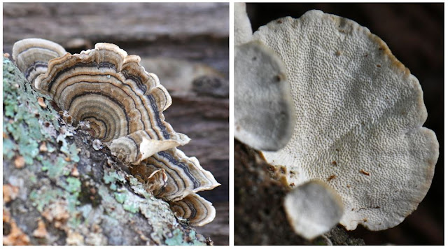
| Turkey
Tail bracket fungus Striped top surface (left); white bottom surface with visible pores (right) |
Turkey Tail and False Turkey Tail (below) bracket fungi are two of the most commonly observed wood-rotting fungi in the woods around Athens. They persist on dead hardwood long after they have released their spores because of their tough structure. The banding on the top surfaces of both species is quite variable, so you have to look underneath to identify them. Turkey Tail is white underneath with visible round pores; False Turkey Tail is smooth underneath, with no visible pores, and tan-colored. Turkey Tail is widely sold for its medicinal value.
 |
| Gilled
Polypore Top surface (left), bottom surface with gills (right) |
It is highly unusual for a polypore (“many pores”) fungus to have gills – but this one does. This species was long placed in a different genus, Lenzites, until DNA studies proved that it is in fact closely related to Turkey Tail.
 |
| Gloeoporus dichrous (no common name) Top surface (left), bottom rubbery surface with pores (right) (Photos by Bill Sheehan) |
Michael Kuo maintains a marvelous website on fungi, www.mushroom.expert.com, that’s both technical and humorous at times. Here’s what he says about this bracket fungus:
“This interesting little polypore [Gloeoporus dichrous] is a decomposer of hardwood logs across the continent. Viewed from above, its creamy to white cap is a bit boring – but its striking underside features an unexpected brown to reddish-brown pore surface. In fact, the pore surface becomes even more interesting if you have descended deep into the mire of myco-geekishness and are willing to pry at it with a dissecting needle or tweezers: the tube layer is rubbery in consistency, and is separable from the cap as a layer. Now that's entertainment.” (Remember to click or tap on an image to zoom in)
 |
| Ganoderma [maybe] sessile |
 |
|
Little Nest
Polypore |
 |
| Cross-section of the Phellinus crust, showing pores |
 |
| Polypore fungus with irregular pores |
|
|
Crust Fungi
Crust fungi is a general term for the sporulating basidiomycete fungi that lie mostly flat against dead wood (some may have a bit of a lip protruding) and usually lack pores. You’ve got to be a log-turner to be a crust nerd because they’re usually found under dead logs on the ground or low on the sides of dead logs. Some are wispy filaments, some are hard and tough, and some have bumps, teeth, or wrinkles. Some are small discs while others may stretch the length of a dead log.
 |
| A beautiful lemon-colored wispy crust, maybe Phlebia subochracea |
Ascomycete Fungi
We found several representatives of ascomycete fungi. This is a large and varied group of fungi that tends to be small so are usually less frequently noticed than basidiomycetes, the other large group of fungi that includes mushrooms, bracket fungi, crust fungi, jelly fungi, and more. The difference between the two groups lies in how spores are produced and arranged.
 |
| Cramp Balls |
Each
little bump on the surface of Cramp Balls covers a tiny chamber that shoots
out spores. As is typical of ascomycetes, the spores are contained in a column-shaped
sac, usually eight to a sac. Annulohypoxylon,
the Cramp Ball genus, was separated from the genus Hypoxylon based on DNA evidence, but also differs from Hypoxylon by the flat rings surrounding
the ‘bumps’ (ostioles) from which the spores are released, and by the black
carbonaceous interior that does not produce bright pigments when potassium
hydroxide is applied. Zoom in on the upper left and far right fruiting bodies
to see the flat ring (annulus) around the openings. Compare to the orange flesh
of true Hypoxylon, below.
 |
| Hypoxylon sp., maybe H. rubiginosum or H. crocopeplum |
Cup Fungi
 |
| Purple
Jellydisc, a
wood-rotting fungus, grows mainly on the trunks and branches of dead Beech
trees, where it often forms large, showy clusters. (Photo by Bill Sheehan) |
 |
|
Blue-stain fungus (Chlorociboria aeruginosa) is most often seen as a distinctive blue-green stain on well decayed oak logs. These tiny, blue-green cup fungi occur infrequently in summer and fall. |
 |
| Orbilia
sp. Some species in the family Orbiliaceae are carnivorous and trap and digest nematodes. |
For more info on nematode-eating fungi, see Else Vellinga’s article on Oyster mushrooms and their relatives: "Fungal Snares and Other Sticky Ends," here.
 |
|
Two unidentified
cup fungi. |
Slime Molds Are Not Fungi
Once considered part of the fungi kingdom, slime molds don’t fit comfortably into any of the five currently recognized kingdoms, but are placed in the Protista kingdom, a sort of grab bag of primitive, single-celled organisms. The visible slime molds we most often encounter are in the group, Myxogastria. When food is abundant, these slime molds exist as single-celled organisms, feeding on microorganisms that live in any type of dead plant material. When food is in short supply, many of these single-celled organisms come together and start streaming as a single body. They then form reproductive bodies – sporocarps – like those seen below, often a tiny spore-containing ball on a slender stalk. These are typically a millimeter or two high so are most easily spotted when they aggregate. (The Dog Vomit Slime is an exception as it can cover a foot or more on wood chips or other dead wood.) The ability of slime molds in the streaming stage to solve problems, such as finding the shortest path through a maze, has led to their being used to plan rail and subway lines. Some researchers use the term “intelligence” to describe this ability.
 |
| Wasp Nest Slime Mold |
This slime mold has clusters of spherical capsules on short stalks. The spores are borne on filaments within the capsules. Spores are dispersed when the head bursts, leaving the filaments (red fluff in the photo). The empty bottoms of the capsules look like the nests of paper wasps, hence the common name. The scientific name, vesparium, also refers to the family that paper wasps are in, Vespidae.
 |
| Jean
Lodge sharing her extensive fungi expertise with Ramblers |
OBSERVED SPECIES:
Maple Tarspot Rhytisma acerinumChinese Wisteria Wisteria sinensis
Honey Mushroom Armillaria mellea
Witches’ Butter Tremella mesenterica
Deer Mushroom Pluteus cervinus
Leaf-rotting fungus, no common name Gymnopus sp.
False Turkey Tail Stereum ostrea
Gilled Polypore Trametes [Lenzites] betulinus
Wood-rotting polypore, no common name Gloeoporus dichrous
Thin-walled Maze Polypore Daedaleopsis confragosa
Polypore, no common name Ganoderma [maybe] sessile
Little Nest Polypore Poronidulus [Trametes] conchifer
Poroid crust, no common name Phellinus sp.
Polypore fungus with irregular pores unknown species
Cramp Balls Annulohypoxylon sp.
Blue-stain fungus Chlorociboria aeruginosa
Two species of cup fungi unknown species
Slime mold, no common name Physarum sp., likely stellatum
Wasp Nest Slime Mold Metatrichia vesparium
Postscript: the best trail map for the Botanical Garden I've seen.


